Explore Our Exhibits
-
![]()
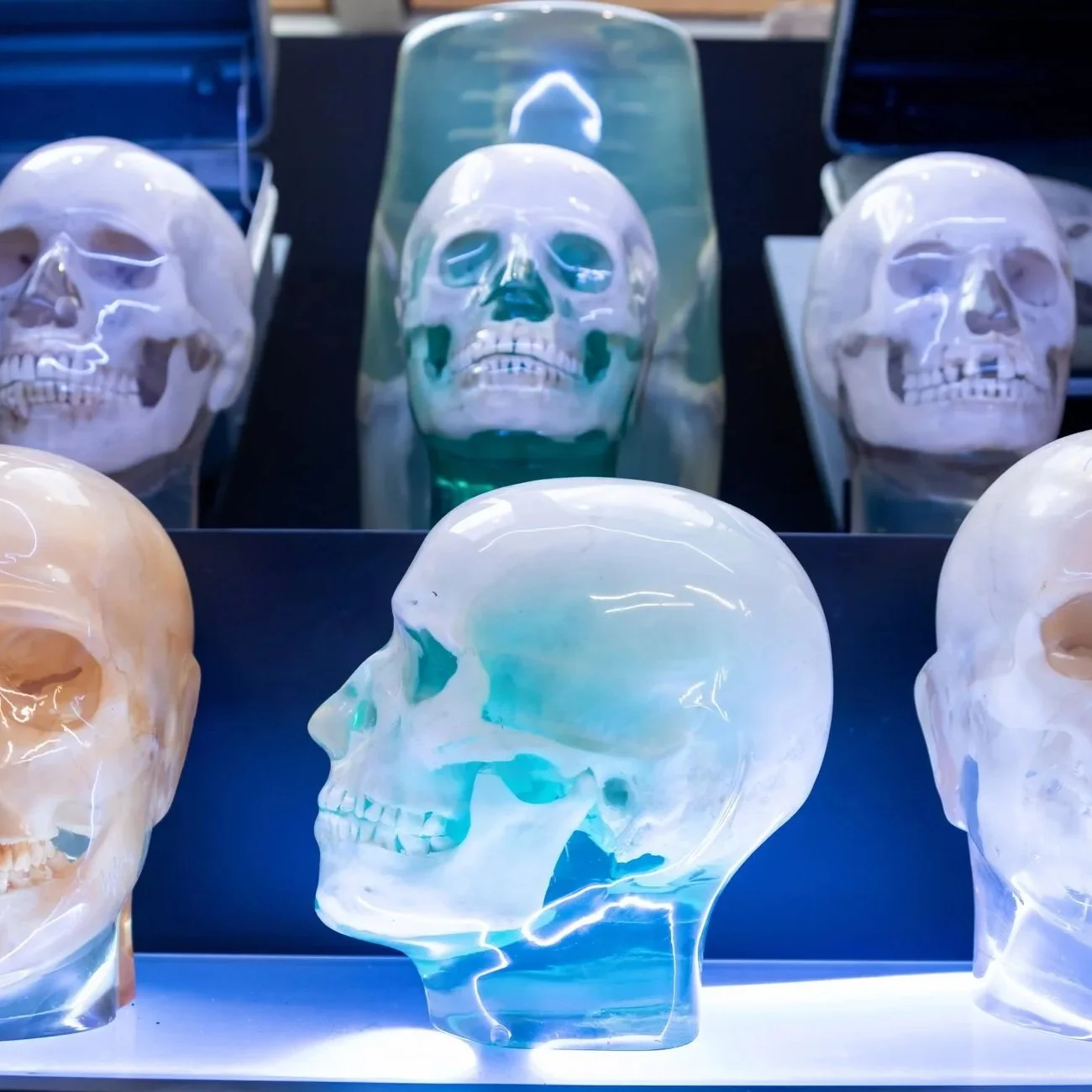
X-Ray Phantoms
X-ray phantoms are specialized tools used in radiography training to teach technologists proper patient positioning, exposure, and image interpretation without subjecting living patients to unnecessary radiation. Historically, these objects served as stand-ins for the human body, allowing repeated imaging under controlled conditions.
Until the 1970s, many x-ray phantoms were constructed using real human bone, embedded in Lucite, a durable, transparent resin that allowed technologists to visualize skeletal structures clearly while withstanding repeated imaging. These phantoms closely replicated true anatomical density and variation, making them especially valuable for training and early imaging research.
As material science evolved and plastic took over real biological specimens, the use of human bone was phased out in favor of synthetic substitutes. While phantoms remain a critical component of radiologic education today, examples made with authentic human bone are now rare.
-
![]()
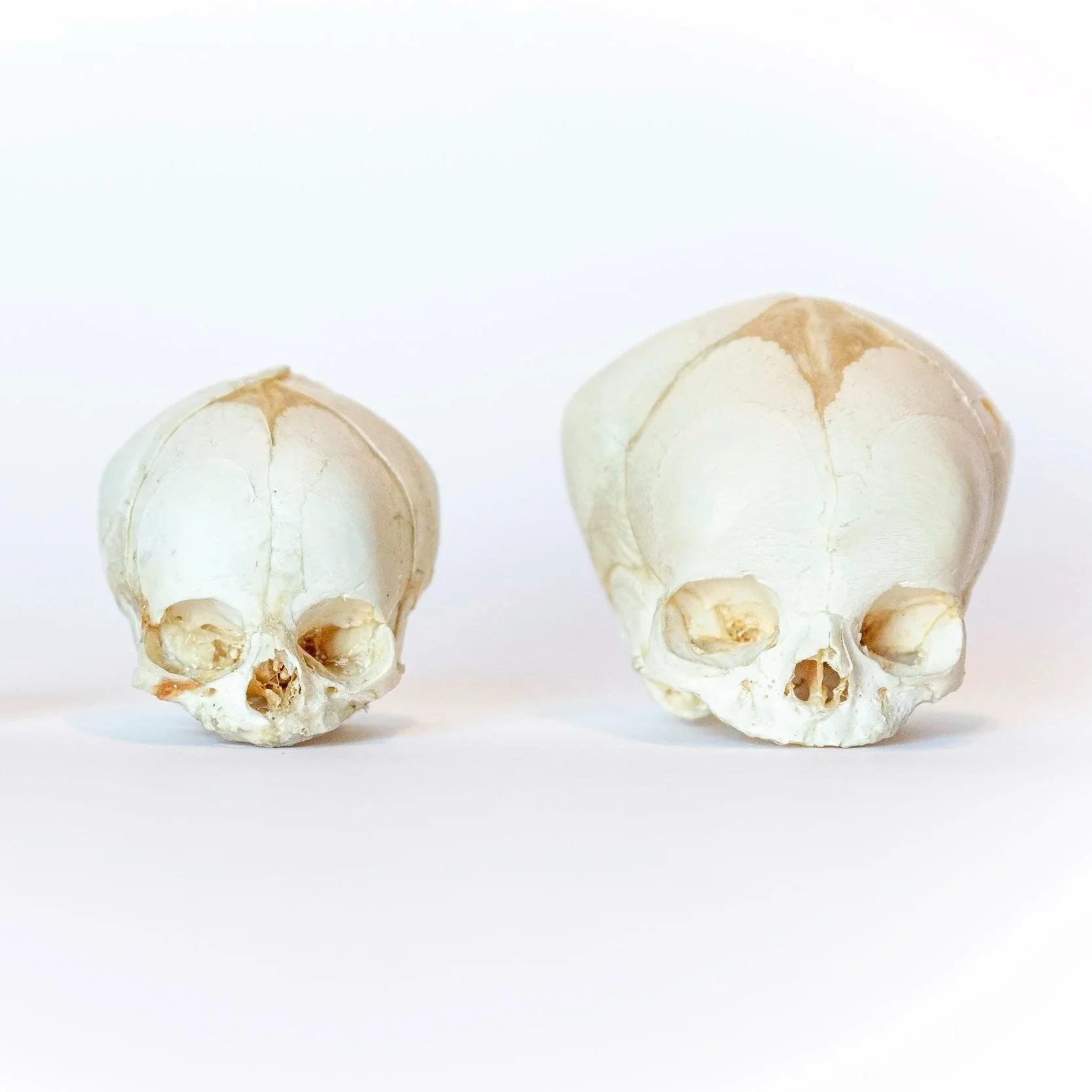
Pediatric Development
This exhibit traces human skeletal development from the fetal period through childhood, adolescence, and into adulthood. The exhibit presents a comprehensive view of both cranial and skeletal development across multiple stages of life.
The cranial component focuses on the formation and gradual fusion of skull plates, demonstrating changes in sutures, fontanelles, facial proportions, and jaw development as the brain and face grow.
The skeletal component examines longitudinal bone growth through ossification centers and epiphyseal (growth) plates, illustrating how different regions of the skeleton mature at different rates.
-
![]()
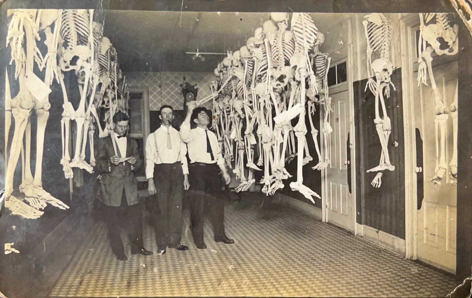
History of the Medical Bone Trade
The Bone Museum houses the most comprehensive database dedicated to the history of the medical bone trade. This exhibit traces the global movement, use, and reuse of medical skeletons from the late 19th century through the 1980s, a period when real human remains were central to medical education and research.
The exhibit stretches throughout the museum, and features original historical materials, such as medical bone boxes, purchase receipts, catalogs, and hundreds of archival photographs.
Together, these objects document how skeletons were prepared, sold, transported, studied, and passed between institutions, schools, and companies across decades
-
![]()
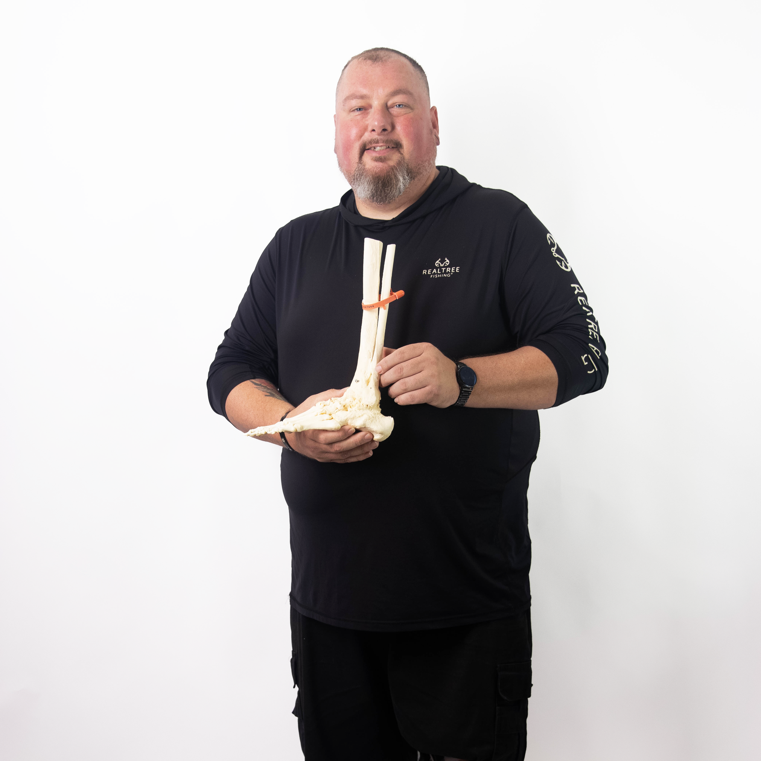
Johnny's Leg
In 2025, the Bone Museum was honored to display the skeletonized leg of a man who underwent a below-the-knee amputation. Johnny was born with a congenital club foot in the 1980s and underwent multiple corrective surgeries throughout his life. Despite extensive intervention, these procedures failed to relieve his pain or restore functional use of the limb.
In 2015, after years of chronic pain and limited mobility, Johnny elected to undergo a below-the-knee amputation. Following the procedure, he chose to have his leg skeletonized and preserved. In 2025, ten years after his amputation, Johnny made the decision to loan his leg to the Bone Museum so that his experience could serve an educational purpose.
This exhibit documents not only the anatomical outcome of long-term surgical intervention and amputation, but also the human story behind it. Johnny’s leg stands as a powerful reminder of the importance of patient autonomy, and medical self-advocacy.
-
![]()
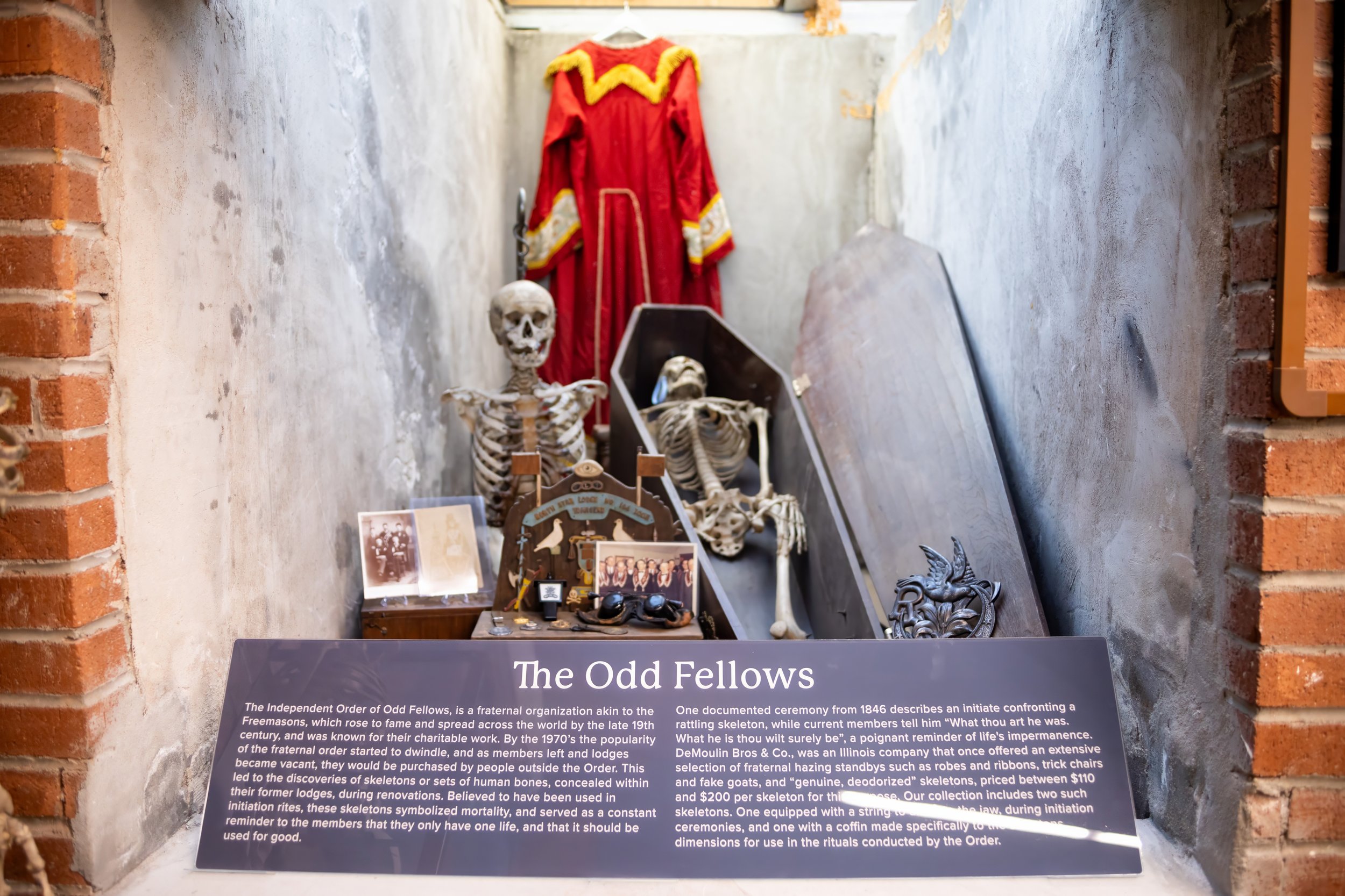
The OddFellows
Medical skeletons have a long and complex history of use beyond the classroom and hospital. In the late 19th and early 20th centuries, real human skeletons were frequently incorporated into the initiations, and symbolic practices of secret societies and fraternal organizations, most notably the Independent Order of Odd Fellows.
Within these spaces, skeletons were used as moral symbols, representations of mortality, secrecy, and transformation, rather than strictly anatomical teaching tools. Their presence reinforced ritual drama and underscored themes of enlightenment, rebirth, and the fragility of life.
The practice became so widespread that medical supply companies openly catered to this demand. Anatomical catalogs of the period often included dedicated sections labeled “Human Bric-A-Brac,” offering skeletons and skeletal parts specifically marketed for non-medical use.
-
![]()
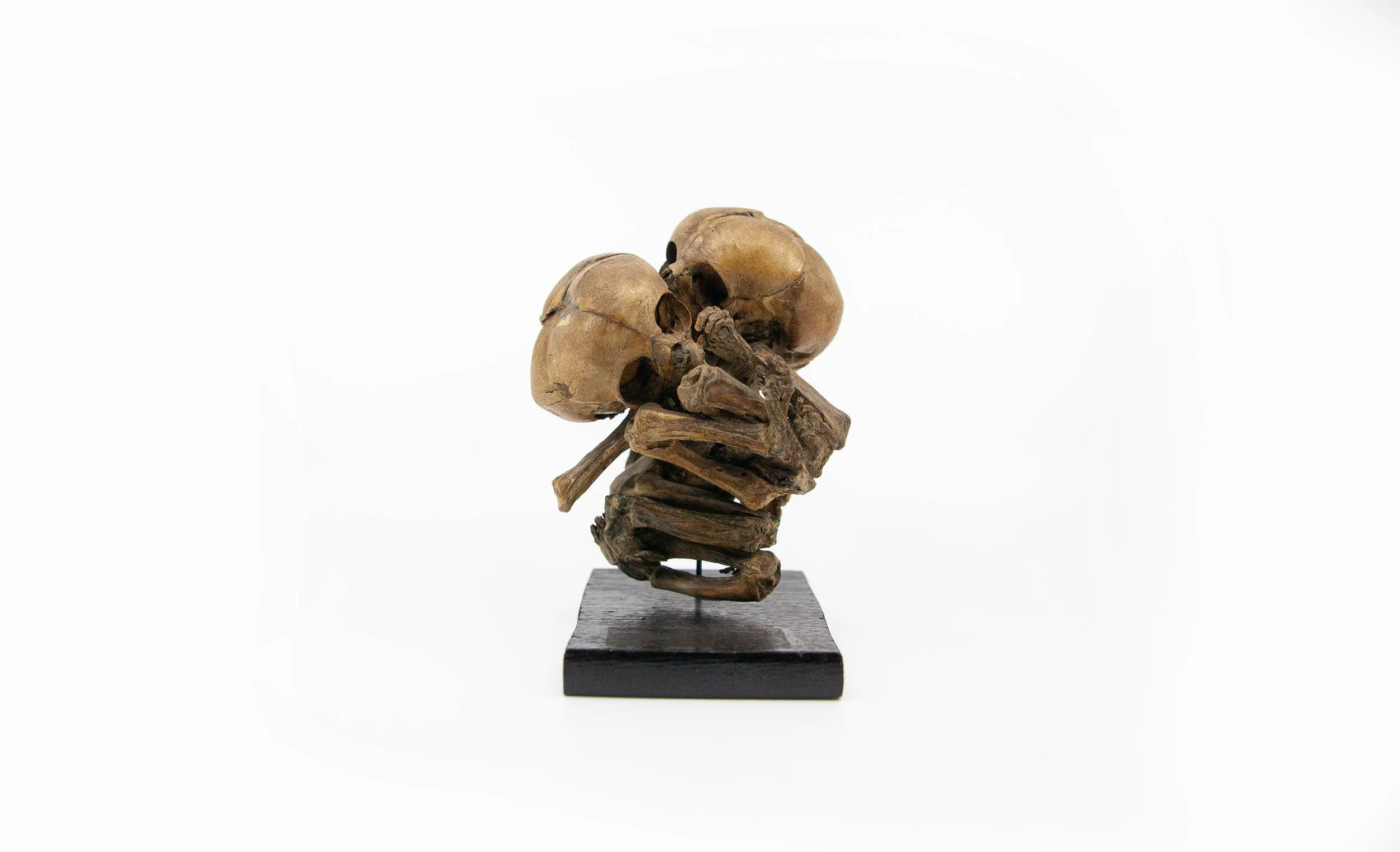
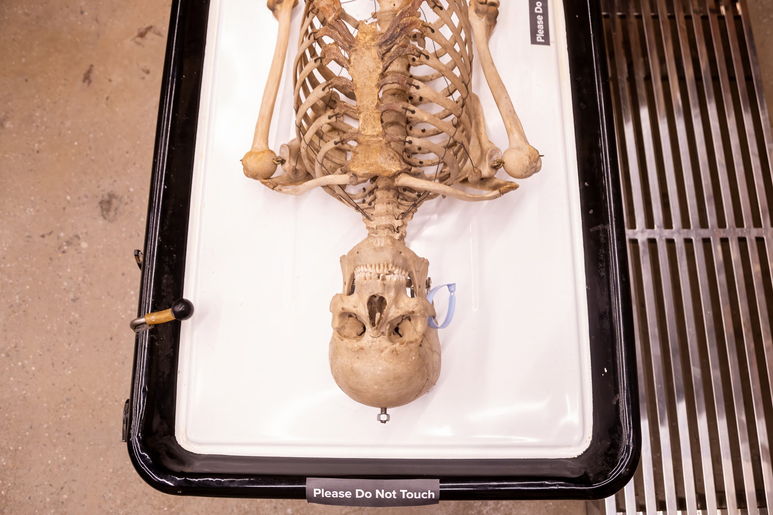
Skeletal Pathologies and Trauma
The Bone Museum houses an extensive exhibit dedicated to skeletal pathology and trauma, showcasing how disease, injury, and medical intervention leave lasting evidence in bone. This collection includes rare and significant specimens such as conjoined twins, advanced skeletal changes associated with tertiary syphilis, and developmental conditions including craniosynostosis and microcephaly.
The exhibit also examines traumatic injury and healing, featuring examples of severe fractures such as complete femoral breaks with non-union and malunion, illustrating both failed and altered healing processes. Surgical intervention is represented through historical craniotomies and Le Fort fracture patterns, offering insight into the evolution of trauma care and surgical techniques.
-
![]()
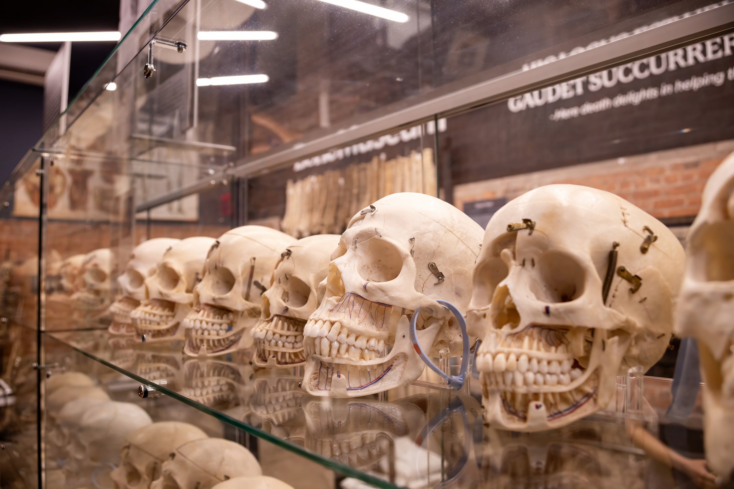
Bone Museum Skull Collection
The Bone Museum houses a collection of over 200 human skulls, representing a wide range of medical preparation styles and educational uses. The collection includes typical medical skulls, explanatory skulls, and demonstrative skulls, each prepared to emphasize different aspects of cranial anatomy and anatomical instruction.
Alongside preparation techniques, the collection documents pathological and traumatic changes to the skull. These include examples of progressive tooth loss, dental abscesses, and other conditions that reveal how disease, injury, and aging alter cranial and dental structures over time.
-
![]()
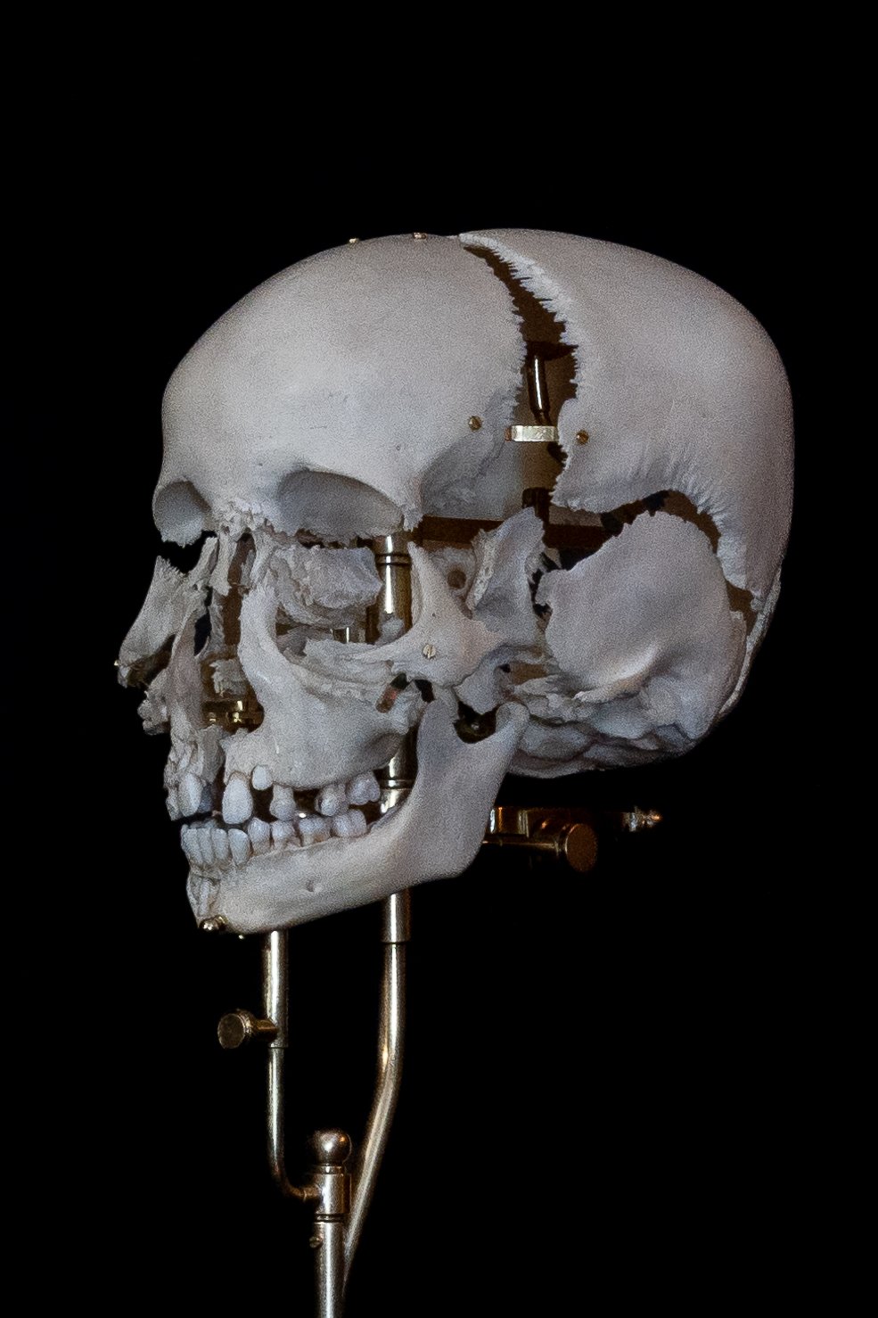
Beauchene Skulls
Beauchêne skulls, commonly referred to as “exploded skulls,” is a distinctive anatomical preparation technique designed to reveal the complex three-dimensional structure of the human cranium. In these preparations, the bones of the skull are carefully separated and mounted to demonstrate their individual forms, points of articulation, and spatial relationships.
The exhibit showcases a range of preparation styles, illustrating different methods of suspension, spacing, and movement. Some skulls are designed to open or expand in specific directions, while others allow bones to pivot or separate along particular anatomical planes. These variations reflect different educational goals, such as studying varying cranial thickness, or sutural relationship.
-
![]()
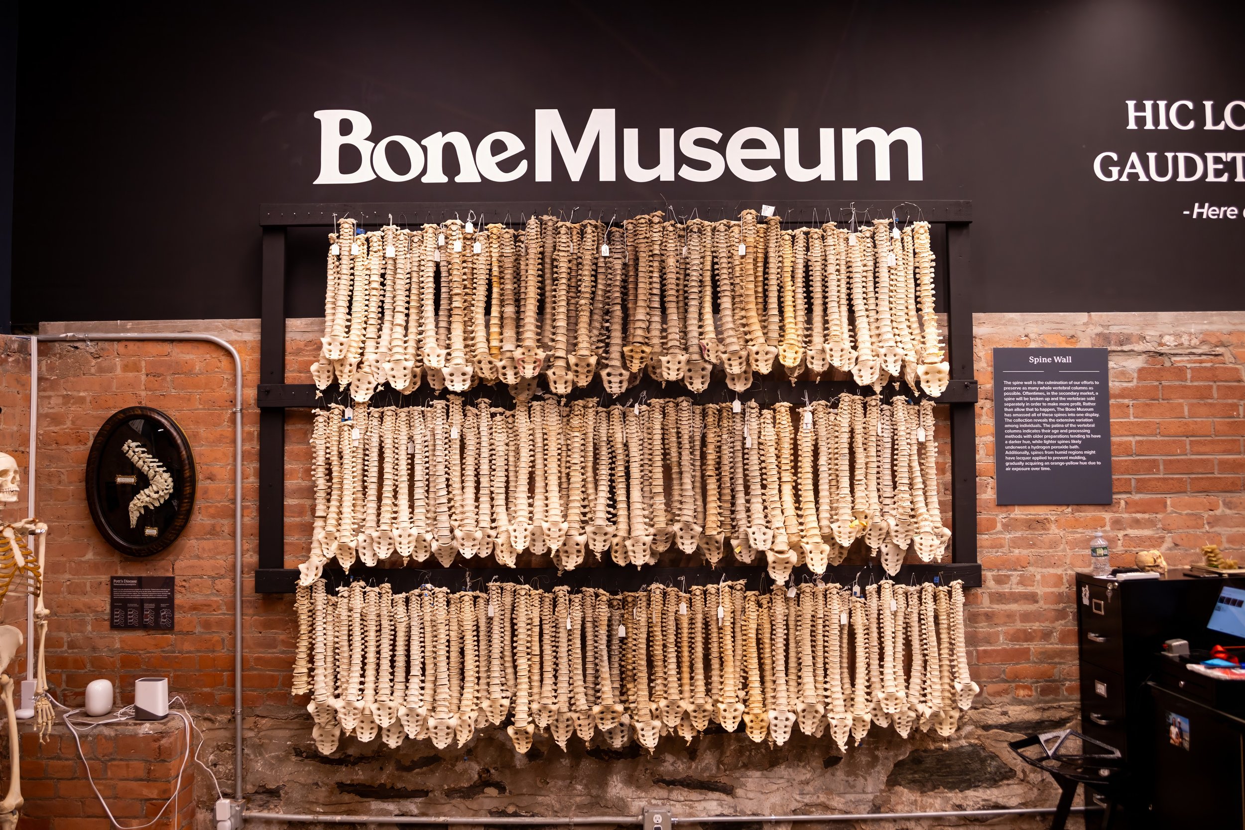
The Spine Wall
The Bone Museum hosts the largest publicly available collection of real human spinal columns, colloquially known as the “Spine Wall”. This collection features over 150 spines with varying pathologies and historical preparations and backgrounds.
The Spine Wall is a comparative research and educational resource, allowing for close examination of vertebral morphology, curvature, fusion patterns, and degenerative change.
Included in the exhibit are spines with indiopatihc and pathological changes such as scoliosis, and spinal tiberculosis.